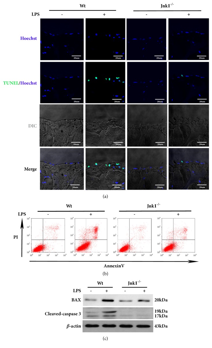
Figure 5.
JNK1 knockout decreases peritoneal mesothelial cells apoptosis induced by LPS. (a) Representative images of TUNEL staining in parietal peritoneum of Wt and Jnk1-/- mice after LPS treatment. Scale bar: 20um. (b) Peritoneal mesothelial cells derived from Wt or Jnk1-/- mice and exposed to LPS, cell apoptosis was examined by flow cytometry. (c) Cell lysates were probed with antibodies against BAX and cleaved-caspase 3.

