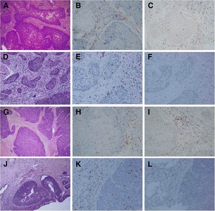
Fig. 1.
CD8+ and Foxp3+ T cells in cervical cancer before and after NACT. A pre-chemotherapy biopsy showed a typical morphology of squamous cell carcinoma (a). The tumor had higher CD8+ T cells (b) and Foxp3+ cells (c) in the surrounding tissue than those in the tumor nests. After NACT, the tumor didn’t achieve pathological complete response, which harbors sparse tumor nests in the stroma (d). Foxp3+ T cells decreased significantly (e), while CD8+ cells remained stable (f). In another case with pathological complete response, pre-chemotherapy biopsy was characteristic of squamous cell carcinoma (g). Both CD8+ T cells (h) and Foxp3+ cells (i) were higher in the surrounding tissue than those in the tumor nests. After NACT, the tumor only had a component of residue carcinoma in situ (j). CD8+ cells were not significantly changed (k), while Foxp3+ T cells were almost undetectable (l)

