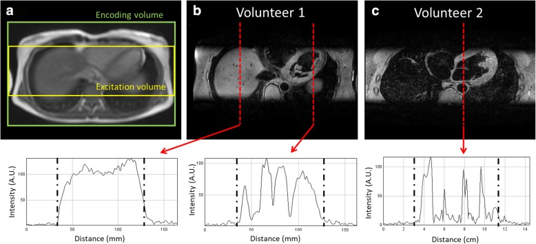
Fig. 3.
Scan planning of 3D FSE in coronal orientation, using a narrower excitation volume than encoding volume in the slice direction (a) to spatially encode potential artifacts arising from the non-selective refocusing pulses. Resulting 3D FSE image for two healthy subjects, demonstrating excellent suppression of outer volume tissue (b and c). Signal intensity profiles through the liver and heart highlight the difference in signal between tissue within the excited volume and outer volume which are separated in the plots by the dashed lines

