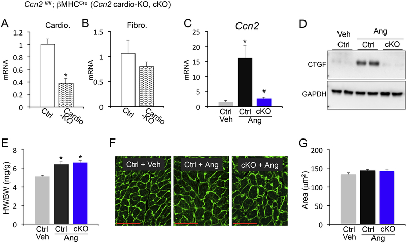
Figure 1 – Analysis of cardiomyocyte-specific CCN2 deletion mice.
(A, B) qPCR analysis for Ccn2 mRNA expression normalized to Rpl7 housekeeping gene in isolated cardiomyocytes (A) or isolated cardiac fibroblasts (B) from the indicated genotypes. (C) qPCR analysis for Ccn2 mRNA expression normalized to Rpl7 housekeeping gene in hearts from vehicle (veh) treated control (ctrl) mice or angiotensin II (Ang) stimulated control (ctrl) or Ccn2 cardiac KO mice (cKO). n≥ 3 biological replicates / condition. (D) Western blot for CCN2 and GAPDH loading control from cardiac protein extracts from the indicated genotypes and treatments. (E) Heart weight to body weight ratio (HW/BW) from the indicated groups of mice. n≥ 5 biological replicates / condition. (F) Representative Wheat Germ Agglutinin (WGA) staining outlining cell borders (green) in cardiac cross-sections from vehicle and angiotensin II stimulated control and Ccn2 cKO mice. Scale bar is 50μm. (G) Quantification of cardiomyocyte cross-sectional areas from WGA stained sections using ImageJ NIH software. n≥3 hearts / condition, n=100 cells / heart. *p<0.05 vs. ctrl (A) or veh ctrl (C and D); #p<0.05 vs. ctrl ang treatment

