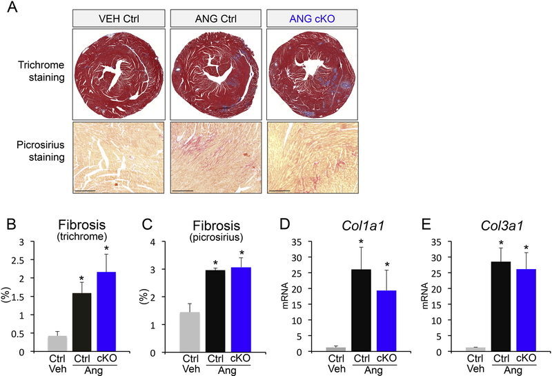
Figure 2 – Cardiomyocyte-derived CCN2 is dispensable for fibrotic remodeling.
(A) Representative Masson’s trichrome-stained histological sections for fibrosis (upper panels; blue) and picrosirius staining (bottom panels; red) in control and Ccn2 cKO mouse hearts with or without angiotensin II infusion. (B, C) Quantification of fibrosis from Masson’s trichromestained and picrosirius-stained histological sections of the indicated genotypes and treatments using ImageJ NIH software. Scale bar is 20μm. n≥5 animals / condition (D, E) qPCR analysis for collagen 1a1 (Col1a1) and collagen 3a1 (Col3a1) mRNA expression normalized to Rpl7 housekeeping gene in hearts from the indicated groups. n≥5 biological replicates / condition. *p<0.05 vs. veh ctrl

