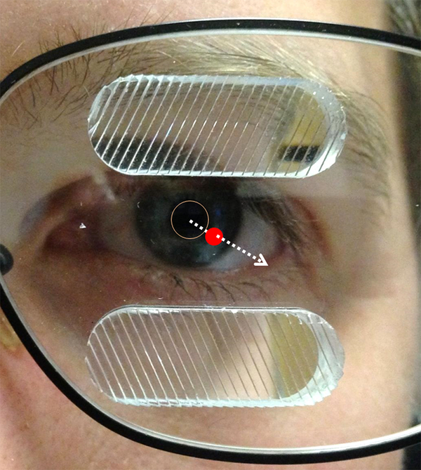
Figure 1.
Standard unilateral fitting of oblique 57Δ peripheral prisms (p-prisms) on the left eye for left homonymous visual field defect (HVFD) with the segments laterally centered on the visual axis in primary gaze (indicated by the dashed arrow). The red dot is the position of the visual axis at the spectacle plane (picture was taken from slightly off to the left and up from the photographer’s perspective). The p-prisms straddle the border of the HVFD such that half of the prism is within the intact (right) visual field when in primary position of gaze.

