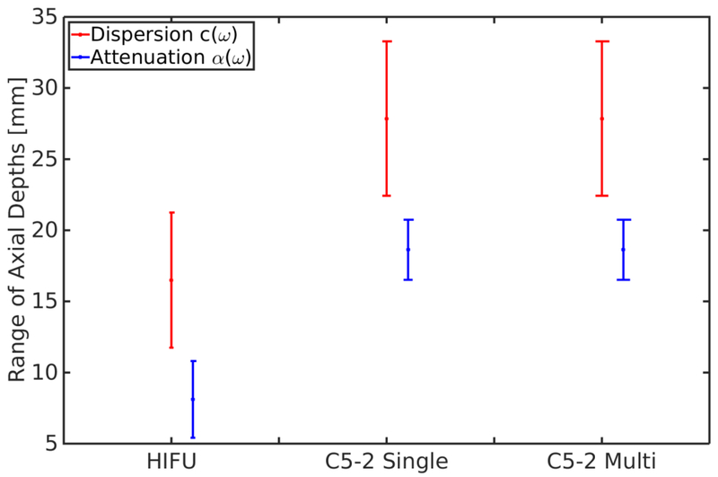
Figure 7:
The range of depths that varied < ± 5% from the dispersion or attenuation estimate at the center of the excitation for the HIFU piston, single focus, and multi-focal C5-2v curvilinear array excitations in the VE material M4 (Table 2). Errorbars represent the variation seen over the frequency range of 100-400 Hz. As the DOF of the excitation increases from F/1 in the HIFU piston to F/2 in the single focus C5-2v excitation the range of depths that give a consistent estimate also increases, although the range of depths for attenuation is always smaller than the range of depths for dispersion. However, the range of depths using a multi-focal excitation did not significantly change relative to its single focus counterpart. The range of depths were asymmetric about the focal depth due to axial asymmetries in the excitation. In general the range of depths tended to skew shallow to the focal depth as seen at 200 Hz in Figure 6. Note that these estimates do not take into account bias in an estimate, only the consistency of the measurement made.

