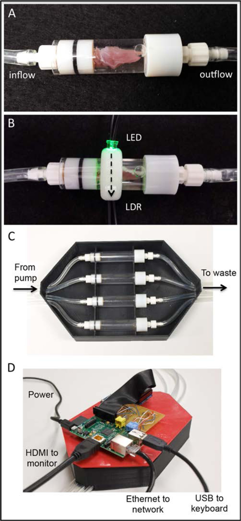
Figure 1:

The primary components of the as built device are the muscle infusion chambers and optical detection collars (A and B). SDS solution is delivered into muscle tissue via a hypodermic needle (not seen), infused through the muscle, and outflows to waste collection. Light produced by the LED travels across the chamber and though the muscle sample (B: dashed arrow) where it is detected and converted to a voltage output by the LDR. Infusion units, optical monitoring collars, and data collection hardware are housed within and mounted onto a custom fabricated enclosure (C and D). The enclosure was designed to accommodate four side-by-side decellularization units each capable of accommodating a single muscle tissue sample. LDR output voltage was converted to a digital signal (8-bit) and stored with the aid of integrated data collection hardware (Raspberry Pi) and software (Python).
