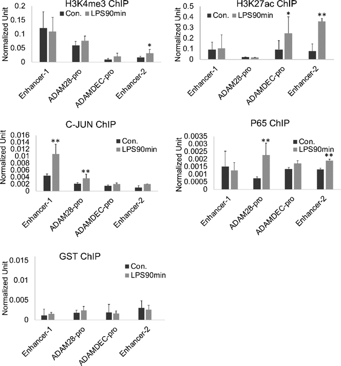
Figure 5. ChIP-seq of the enhancer regions.
MonoMac6 cells were treated with 1μg/ml LPS for 90 minutes. ChIP assays were performed with antibodies H3K4me3, H3K27ac, c-JUN and P65. In resting cells, H3K4me3 was highest at Enhancer 1. LPS stimulation let to increased c-JUN at Enhancer 1, ADAM28 promoter, H3K27ac at the ADAMDEC1 promoter and Enhancer 2 as well as binding of p65 to the ADAM28 promoter and Enhancer 2. (n=4, error bars represent SD, * indicates p<0.05, ** p<0.01).

