
Figure 2.
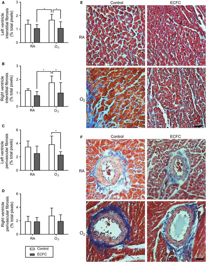
Effect of ECFC treatment on the left and right ventricle interstitial and perivascular fibrosis. (A) Left ventricle and (B) right ventricle interstitial tissue fibrosis (% total pixels) and (C) Left ventricle and (D) right ventricle perivascular fibrosis (% total pixels) of 28 days old rats exposed to high concentration of oxygen (O2) versus room air (RA) in the neonatal period and subsequently treated with endothelial colony‐forming cells (ECFC) versus saline (Control). (E) Representative photomicrographs of LV myocardium and (F) LV myocardium vessel stained with Masson's trichrome. Magnification of X400; scale bar, 50 μm. *P < 0.05.