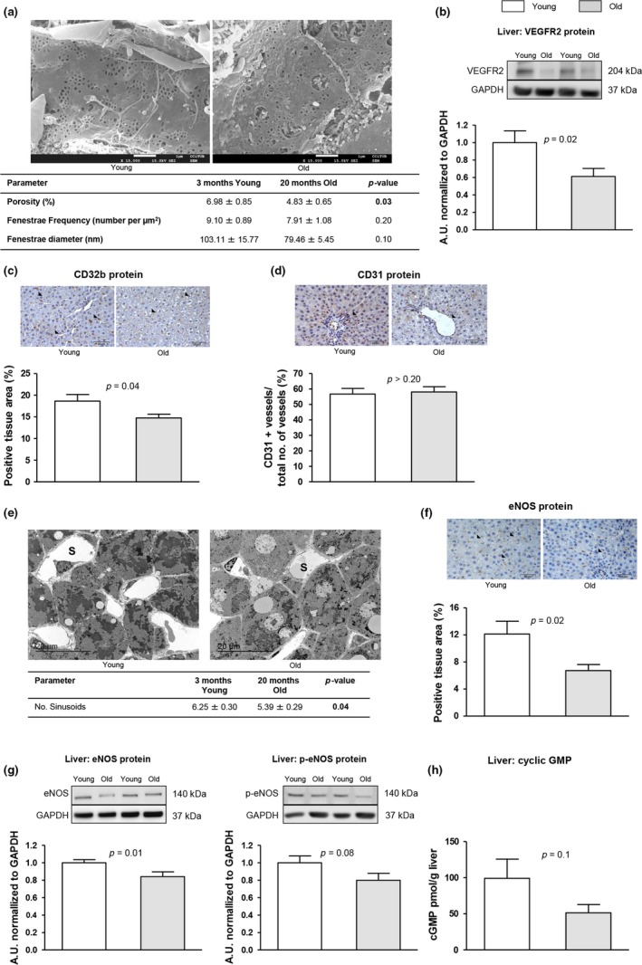
Figure 2.

Endothelial dedifferentiation in the aged liver. The following markers of sinusoidal endothelial phenotype were analyzed in liver tissue from young and aged rats. (a) Representative scanning electron microscopy images and quantification of porosity, fenestration frequency, and fenestration diameter. (b) VEGFR2 protein expression normalized to GAPDH. (c) Representative images of CD32b immunohistochemistry and corresponding quantification. (d) Representative images of CD31 immunohistochemistry and corresponding quantification. (e) Representative transmission electron microscopy images and quantification of numbers of sinusoids. (f) Representative images of eNOS immunohistochemistry and corresponding quantification. (g) Representative western blots of eNOS and p‐eNOS, and protein quantification normalized to GAPDH. (h) Levels of cyclic GMP. n = 3 (a and e) and n = 12 (other panels) per group. Results represent mean ± SEM. All images 400x, scale bar=50 μm
