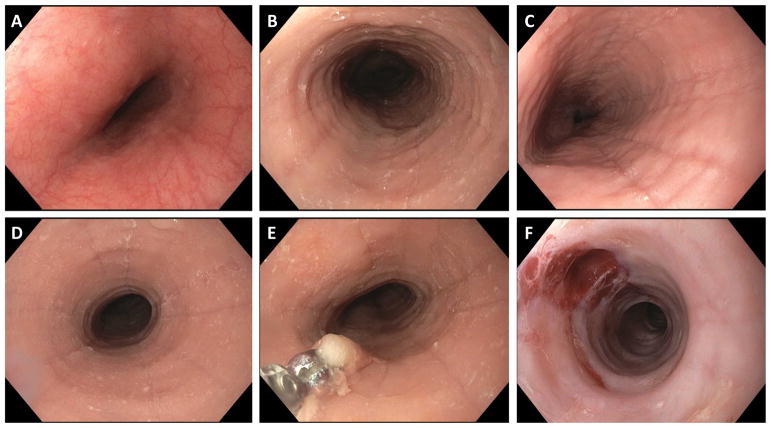
Figure 1.
Endoscopic images of EoE. (A) The endoscopic appearance of the normal esophagus. Note the uniform and smooth appearance of the esophageal mucosa, with the fine vascular pattern clearly visible. (B) An EoE patient with evidence of esophageal rings, furrows, edema, and exudates. (C) An EoE patient with esophageal edema, deep furrows, and mild exudates. (D) An EoE patient with a focal stricture, in addition to mild rings, furrows, edema, and exudates. (E) Esophageal biopsy underway. (F) An EoE patient with a very narrow caliber esophagus and tight rings, as well as edema, after esophageal dilation. Good dilation effect (mucosal rent) is seen in the 11 o’clock position.

