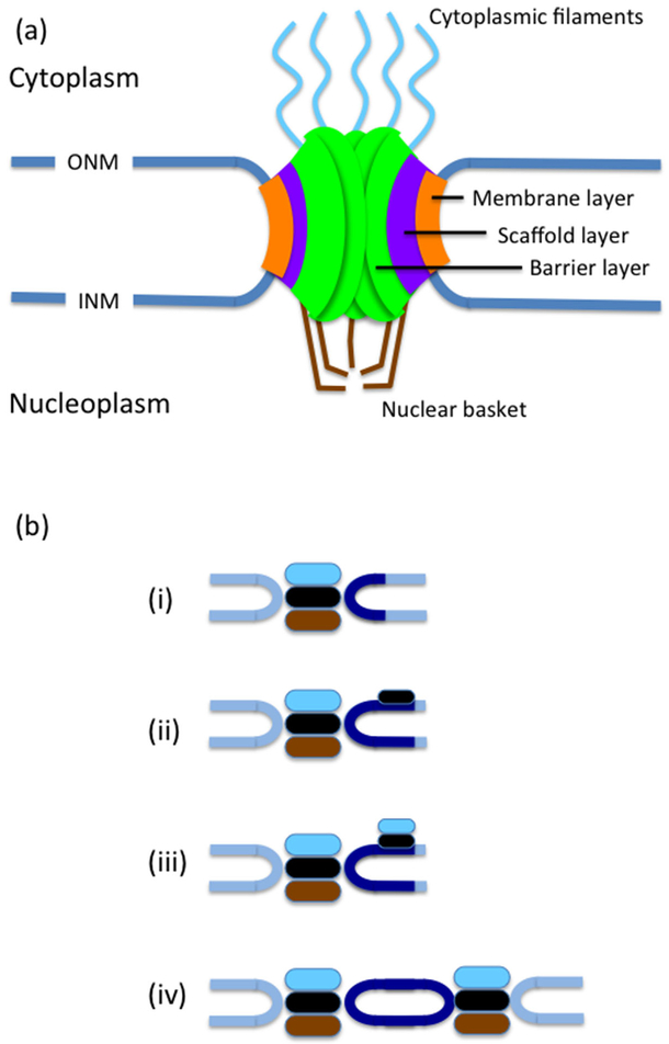
Fig. 4.
The NPC and SPB. (a) A simplified diagram of the NPC, showing the membrane layer embedded in the membrane connecting the outer and inner nuclear membranes, the scaffold layer and the barrier layer. In addition the NPC has a nuclear basket and cytoplasmic filaments. (b) Stages of SPB insertion: (i) The SPB is composed of 3 plaques, an outer plaque (light blue), central plaque (black) and inner plaque (brown). Next to the SPB is the half bridge (dark blue). (ii) The half bridge extends and the satellite assembles at its distal end.(iii) Outer plaque components are added to the satellite. (iv) The new SPB is inserted into the NE and components of the inner plaque are added.

