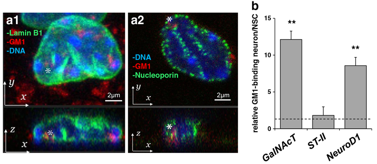
Fig. 3.
Nuclear GM1 in at the nuclear periphery and is accumulated in the activated GalNAcT and NeuroD1 genes in neurons. (a) Localization of GM1 at the nuclear periphery in neurons. Differentiated neurons were fixed, stained with fluorescent cholera toxin B subunit (CtxB, red fluorescent) to determine the localization of GM1. Cells were co-stained with lamin B1 (green fluorescence in a1) or nucleoporin (green fluorescence in a2). Nuclear DNA was counterstained with Hoechst 33,258. Z-projection: bottom panel is x-z plane. (b) GM1 is accumulated in the activated GalNAcT and NeuroD1 genes. The nuclear fraction of differentiated neurons with prior formaldehyde cross-linking was used for anti-GM1 immunoprecipitation. The amount of specific DNA fragments co-precipitated with GM1 was analyzed by quantitative real-time PCR. The data indicate the relative GM1 binding ability in neurons. The value of NSC samples is defined as 1.0 and represented with a dashed line. Each bar represents mean ± SD of 3 to 4 independent experiments (n = 3–4). ** (p < 0.01) indicates the level of significance in two-tailed t-tests of differences between differentiated neurons versus NSCs

