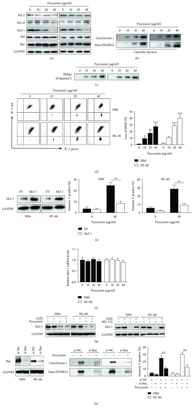
Figure 3.
Camalexin triggers the mitochondrial dysfunction pathway. (a, b, c) NB4 and HL-60 cells were treated with indicated doses of camalexin, and then total cellular lysates were subjected to Western blot analysis with indicated antibodies. Activation of Bax was assessed by immunoprecipitation using active conformation-specific antibody. The cytosolic fractions of cells were subjected to Western blot analysis. (d) NB4 and HL-60 cells were exposed to various doses of camalexin for 6 h; disruption of MMP was indicated by the increase of proportion of cells with green fluorescent (left) and decrease in the proportion of cells with higher red (JC-1 aggregates)/green (JC-1 monomers) ratio of JC-1 fluorescent (right). (e) NB4 and HL-60 cells were exposed to various doses of camalexin for 12 h, and then Mcl-1 mRNA levels were evaluated by RT-PCR. (f) NB4 and HL-60 cells were treated with CHX (100 μM) or MG132 (300 nM) in the presence or absence of camalexin (40 μM) for 24 h, and then total cellular lysates were subjected to the Western blotting analysis. (g) Cells were transfected with empty vector (EV) or Mcl-1; the expression of Mcl-1 was analyzed by Western blotting (left). After transfection for 24 h, cells were treated with or without camalexin (40 μM) for another 24 h, and then cellular apoptosis was assayed by flow cytometry (center and right). (h) NB4 and HL-60 cells were transfected with siRNA against Bax for 24 h, and then the expression levels of Bax were evaluated by Western blotting (left). Then, cells were treated with or without camalexin (40 μM) for another 24 h and the release of cytochrome c and Smac/DIABLO into cytosol was measured by Western blot (center). The cellular apoptosis was assayed by flow cytometry (right). Mean and SD of three independent experiments performed in triplicate are shown; ∗p < 0.05, ∗∗p < 0.01, and ∗∗∗p < 0.001.

