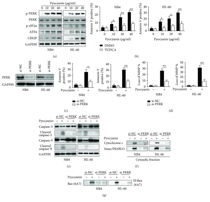
Figure 4.
Camalexin induces ER stress in leukemia cells. (a) NB4 and HL-60 cells were treated with various doses of camalexin for 24 h, and then total cellular lysates were subjected to Western blot analysis with indicated antibodies. (b) NB4 and HL-60 cells were treated with various doses of camalexin in the presence of TUDCA (20 μM) or not for 24 h, and then cellular apoptosis was assayed by flow cytometry. (c) NB4 and HL-60 cells were transfected with si-PERK for 24 h, and then PERK levels were evaluated by Western blotting. (d) NB4 and HL-60 cells were transfected with si-PERK for 24 h, and then cells were treated with camalexin (40 μM) for another 24 h and cellular apoptosis was measured by flow cytometry. (e) NB4 and HL-60 cells were transfected with si-PERK for 24 h, and then cells were treated with camalexin (40 μM) for another 24 h and disruption of MMP was measured. (f, g) NB4 and HL-60 cells were transfected with si-PERK for 24 h, and then cells were treated with camalexin (40 μM) for another 24 h, and then total cellular lysates, cytosolic fractions, and activation of Bax were subjected to Western blotting analysis. Mean and SD of three independent experiments performed in triplicate are shown; ∗p < 0.05 and ∗∗p < 0.01.

