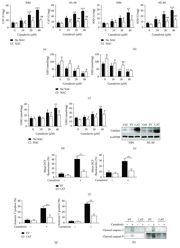
Figure 6.
Camalexin-induced oxidative stress is essential for apoptosis. (a, b, c, d) NB4 and HL-60 cells were treated with various doses of camalexin with or without NAC (20 μM), and then the activities of CAT and SOD and the levels of GSH and GSSG were assayed. (e) NB4 and HL-60 cells were transfected with empty vector (EV) or human catalase (CAT) for 24 h, and then the protein levels of CAT were measured by Western blotting. (f) NB4 and HL-60 cells were transfected with empty vector (EV) or human catalase (CAT) for 24 h, and then cells were exposed to camalexin (40 μM) for 12 h, and then cellular ROS levels were measured by flow cytometry. (g, h) NB4 and HL-60 cells were transfected with empty vector (EV) or human catalase (CAT) for 24 h, and then cells were exposed to camalexin (40 μM) for 24 h, and then cellular apoptosis was measured and lysates were subjected to Western blotting analysis with indicated antibodies. Mean and SD of three independent experiments performed in triplicate are shown; ∗, #p < 0.05, ∗∗, ##p < 0.01, and ∗∗∗p < 0.001.

