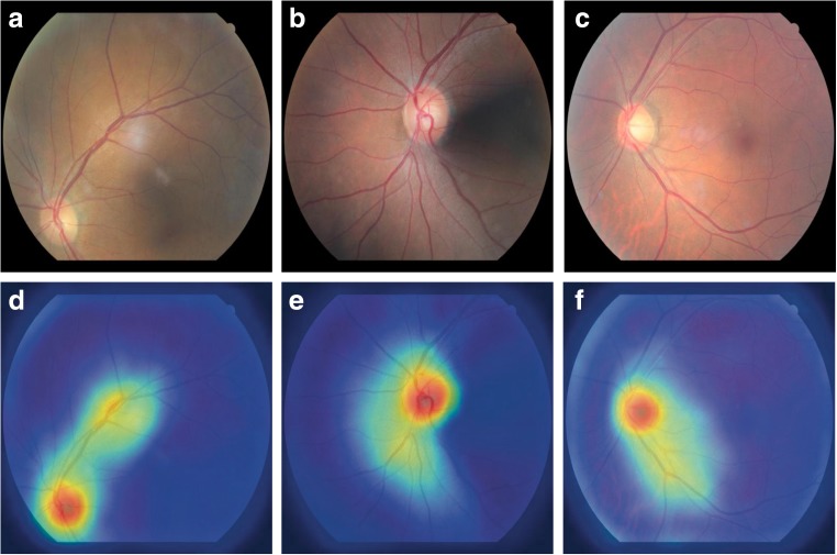
Fig. 3.
Guided Grad-CAM activation maps generated from superior, nasal, and center fundus images. a–c are original fundus images (superior, nasal, center) and d–f correspond to activation maps of superior, nasal, and center fundus, respectively. High activations are observed at the location of optic disc and the surrounding vasculature

