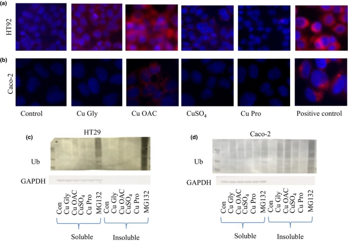
Figure 10.

Fluorescent aggresome images of HT29 (a) and Caco‐2 (b) after a 2‐hr exposure and 24‐ and 10‐hr recovery, respectively. The aggresome detection reagent shown in red is present close to nucleus (stained with Hoechst). Images were combined with MetaMorph and are representative of at least five replicates for each cell line. MG132 was used as a positive control that inhibits proteosomal activity. (c) and (d) show the presence of ubiquitinated proteins in HT29 and Caco‐2 cells, respectively, in the soluble and insoluble fraction (contains aggregated proteins). MG132 shows strong staining especially in the insoluble fraction. GAPDH was used a loading control, and the lack of signal in the insoluble fraction indicates good separation
