
Fig. 1. Crystal structures of human σ1 receptor bound to the classical antagonists haloperidol and NE-100.
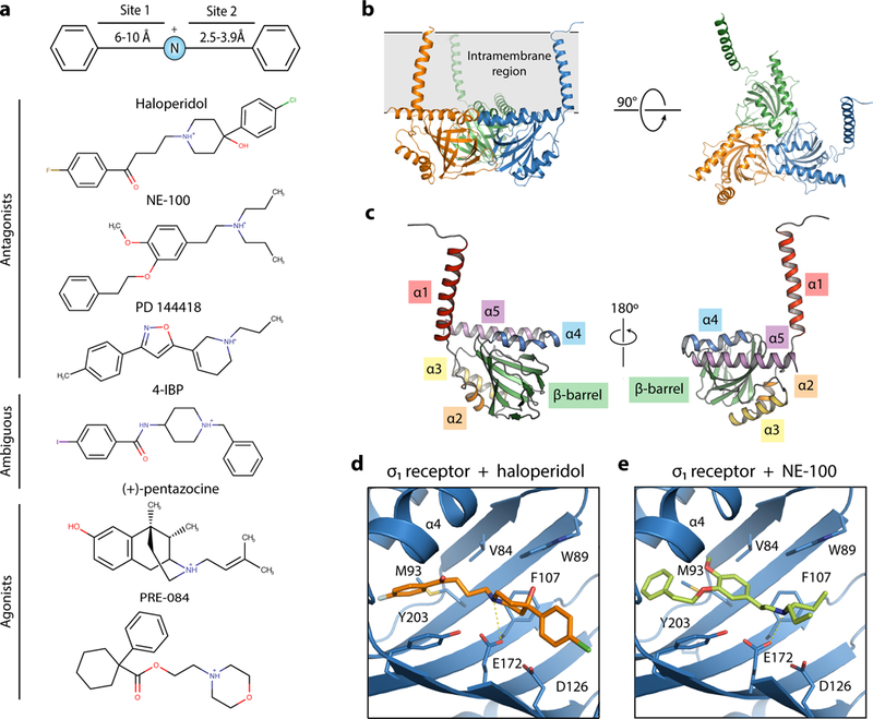
| a, σ1 ligand pharmacophore, based on the work of Glennon et al.8 Representative σ1 ligands are shown below. b, The overall structure of the human σ1 receptor (PDB 5HK1). c, The structure of a single σ1 monomer, with the secondary structural elements labeled. d and e, The binding pocket of the human σ1 receptor (blue) binding in complex with haloperidol (orange, PDB ID: 6DJZ) (d) and NE-100 (light green, PDB ID: 6DK0) (e).