
Fig. 4. | Molecular dynamics simulation reveals a putative binding pathway for (+)-pentazocine.
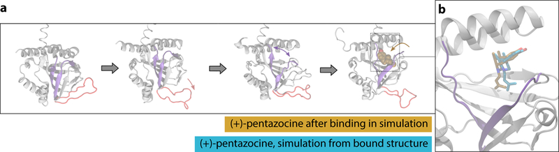
a. Binding pathway of (+)-pentazocine, with the “lid” region shown in red and the beta strands that separate shown in purple. From left to right: simulation begins with an unliganded receptor, where the “lid” region then opens. Next, the interior of the receptor opens, and the ligand enters and binds through this opening. b. A simulation frame after the ligand has entered the binding pocket (tan), compared to a frame initiated from the crystal structure with the ligand bound (blue). Protein backbone is shown for the binding simulation in grey. Helix α4 is located at the top of the rendering.