
Fig. 1 |. Migration of neural-crest-derived progenitors to the primordial gut.
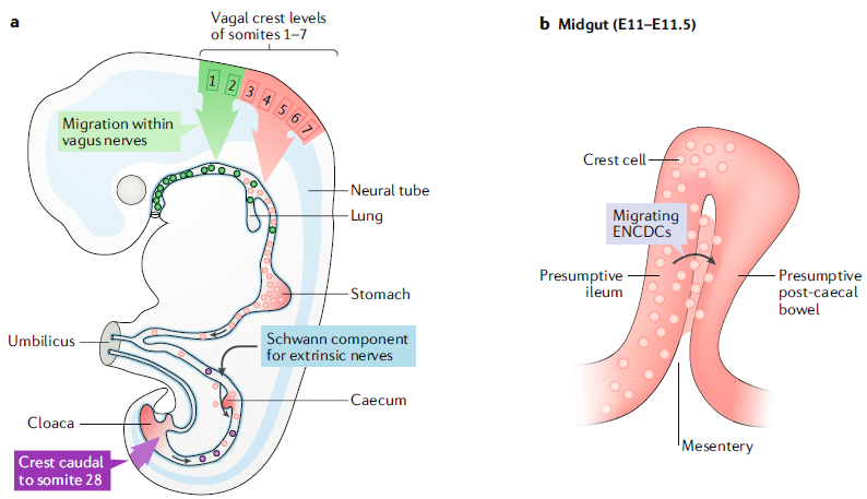
a | A diagram of a developing mouse fetus after the formation of the primordial foregut, midgut and hindgut. The vasculature defines regions of the fetal bowel: the coeliac trunk supplies the foregut, the superior mesenteric artery supplies the midgut and the inferior mesenteric artery, supplies the hindgut. The foregut includes the portion of the bowel from the pharynx to the entry of the pancreatic and bile ducts in the mid-duodenum; the midgut extends to the mid-transverse colon; and the hindgut extends to the ectoderm of the anal canal. The midgut grows rapidly and herniates transiently into the umbilical cord but then folds extensively and rotates upon returning to the abdomen. Neural-crest-derived cells were first shown to migrate to the bowel from the vagal level (corresponding to somites 1–7) and sacral axial level (dark red; caudal to somite 28). A third source of crest-derived precursors of neurons or glia (blue) enter the colon later in ontogeny, among the Schwann cell population found in innervating extrinsic nerve fibres. Most recently, molecular genetic studies have suggested that the vagal level of the crest is more complex than previously suspected. Crest-derived cells from axial levels 1–2 (green) migrate to the oesophagus within the descending fibres of the vagus nerves. Properties of the more caudal vagal crest, at levels 3–7 (red), are more like those of the truncal crest, which gives rise to sympathetic ganglia. This ‘sympatho-enteric’ crest colonizes the entire gut and is the major source of enteric neurons and glia. Sacral-crest-derived cells colonize only the post-umbilical gut. The proportion of enteric neurons derived from the sacral crest (purple) is relatively small and higher distally than proximally. In contrast to vagal-crest-derived cells, which migrate proximo-distally, sacral-crest-derived cells migrate in a distal-to-proximal direction. b | During the folding of the midgut, at embryonic day 11 (E11) to E11.5, the presumptive ileum is transiently located next to a loop of post-caecal bowel (presumptive ascending colon). The dorsal mesentery intervenes between them. A subset of cells within the descending vagal enteric neural-crest-derived cell (ENCDC) population (red) takes a shortcut through the mesentery (pink) to enter the still-to-be colonized gut distal to the caecum. These cells do not have to traverse the caecum. Part a is adapted with permission from REF9, Elsevier.