Figure 7.

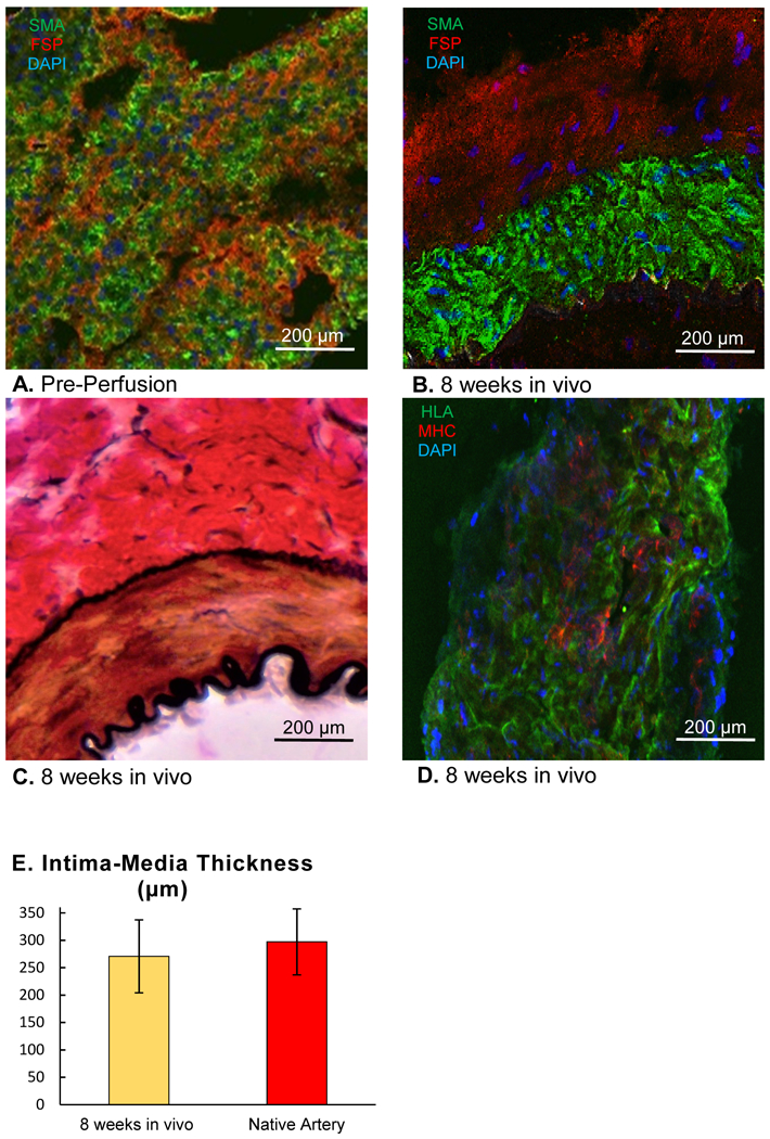
(A) Immunohistochemical staining of a wall segment of the engineered vascular conduit (EVC) immediately after construction (pre-perfusion) with antibodies against smooth muscle actin (SMA, green fluorescent protein) and fibroblast surface protein (FSP, Texas Red), 4′,6-diamidino-2-phenylindole (DAPI): blue. (B) Immunohistochemical staining of a wall segment of the EVC after 8 weeks in vivo with antibodies against smooth muscle actin (SMA, green fluorescent protein) and fibroblast surface protein (FSP, Texas Red), DAPI: blue. (C) Van Gieson staining of a wall segment of the EVC after 8 weeks in vivo. Light Microscopy, 20x. (D) Immunohistochemical staining of a wall segment of the EVC after 8 weeks in vivo with antibodies against human leukocyte antigen A (HLA, green fluorescent protein) and rat-specific major histocompatibility complex I (MHC I, Texas Red). Confocal Microscopy, 20x (B, C, D). (E) Intima-media thickness of EVCs 8 weeks after implantation in vivo compared to that of native femoral arteries, n=5 for both groups. Student’s t-test.
