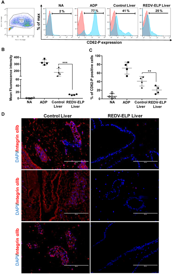
Fig. 6.
Functional analysis of re-endothelialized liver scaffolds. After 3 days of perfusion, liver scaffolds were injected with platelet-rich plasma, incubated for 1 h at 37 °C. (A) Flow cytometry analysis of platelet activation. Platelets were collected from liver scaffolds, and immunostained with CD62-P antibodies. Representative dot plot of platelets population and histograms showing CD62-P expression level of non-activated platelets (NA), platelets stimulated with ADP or collected from unconjugated liver (control) or REDV-ELP conjugated liver. (B) Mean fluorescence intensity of 4 independent experiments showing CD62-P expression level in each sample: NA, ADP, control liver and REDVELP liver (means ± SEM) **P < 0.01 vs. control liver. (C) Percentage of CD62-P positive platelets in each sample: NA, ADP, control liver and REDV-ELP liver (means ± SEM) ***P < 0.001 vs. control liver. (D) Representative immunostaining of re-endohelialized scaffolds using anti-integrin αIIb antibodies (red) and DAPI (blue). Scale bar = 200 lm. (For interpretation of the references to colour in this figure legend, the reader is referred to the web version of this article.)

