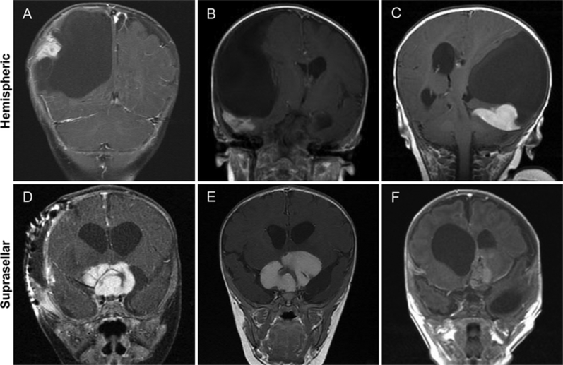
FIG. 1.
A–C: Coronal postcontrast T1-weighted MR images of classically described hemispheric DIGs showing massive cysts with cortically enhancing peripheral nodularity. D–F: Coronal postcontrast T1-weighted MR images of suprasellar DIGs showing large, homogeneously enhancing tumors with small associated cysts.

