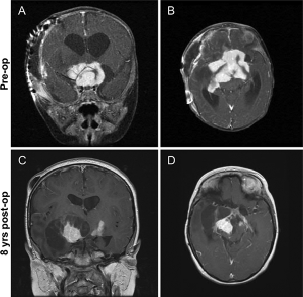
FIG. 2.
Case 1. A and B: Preoperative coronal (A) and axial (B) postcontrast T1-weighted images obtained in a 3-month-old girl with a large, homogeneously enhancing suprasellar tumor, which presented with rapidly enlarging head circumference, nystagmus, and bilateral optic nerve atrophy. The patient underwent subtotal tumor resection and subsequently received chemotherapy and radiation therapy. C and D: Coronal (C) and axial (D) postcontrast T1-weighted images obtained in the same patient 8 years after the first resection, with recurrent tumor growth and cystic transformation. A second partial resection was subsequently performed.

