FIG. 3.
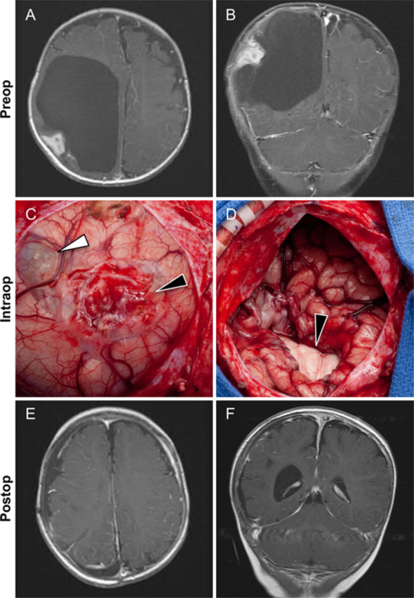
Case 6. A and B: This 4-month-old girl presented with a rapidly enlarging head circumference. Axial (A) and coronal (B) postcontrast T1-weighted images showing a large cystic tumor with an enhancing peripheral cortical-based nodule in the right parietal lobe. C: Intraoperative image obtained in the same patient after opening the dura and wide exposure of right parietal lobe. The solid part of the tumor appears as an extra-axial superficial cortical component (black arrowhead). The cystic portion is also visible (white arrowhead). D: Intraoperative image obtained in the same patient after removal of the tumor. There is significant collapse of the right brain hemisphere. Also seen is the medial wall of the bed of the tumor (black arrowhead). E and F: Axial (E) and coronal (F) postcontrast T1-weighted images obtained in the same patient 1 year after surgery. Neither the cystic or peripheral nodular components are present. Figure is available in color online only.

