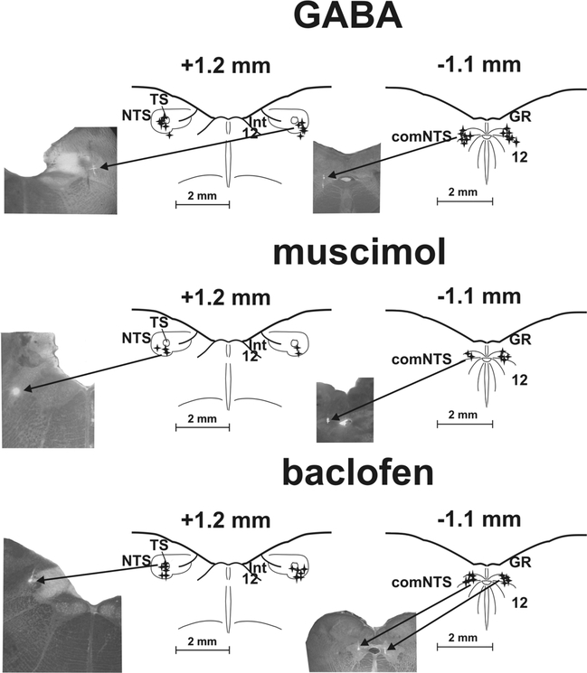
Fig. 1.
Reconstruction of microinjection sites.
Stars represent highlighted positions of the micropipette tip during microinjections of GABA, muscimol and baclofen in the rostral (rNTS; +1.2 mm to the obex) and caudal nucleus of the solitary tract (cNTS; −1.1 mm to the obex) as determined by fluorescent marker. Reference points: comNTS, 12, central canal, medullary surface for the cNTS; TS, NTS, 12, Int, the bottom of the 4th ventricle, medullary surface for the rNTS.
Microinjections of GABA. All 14 microinjection locations were found within or near the commissural subnucleus of the NTS in the cNTS. All 12 microinjections were positioned in or near ventrolateral subnucleus of the NTS in the rNTS.Microinjections of muscimol. All 6 locations where muscimol was delivered in or near comNTS were identified. In the rNTS 5 out of 6 microinjection locations were positively identified in the ventrolateral region of the NTS.
Microinjections of baclofen. All 10 Baclofen microinjections were positioned in or near comNTS in the cNTS. All 12 rNTS microinjection were identified in the area of TS and ventrolateral region of the NTS. 12: hypoglossal ncl., comNTS: commissural subnucleus of the NTS, GR: gracile ncl., Int: ncl. intercalatus, NTS: ncl. tractus solitarius, TS: tractus solitaries
Inset photographs demonstrate the process. The arrows ‘injection’ point out light spots of a spread of fluorescent marker.

