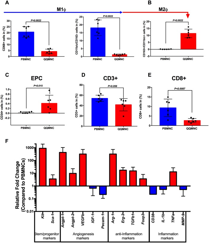
Fig 2. Regeneration-associated cells increased post-QQ culturing.
(A) The flow cytometry analysis revealed that the vast number of PBMNCs were pro-inflammatory monocyte/macrophages (CD68+ and CD11b/c+ /CD163-cells). (B) After QQ culture conditioning, alternatively activated M2ϕ percentages were sharply increased. (C) The proportion of endothelial progenitor cells was higher after QQ culture conditioning. (D) The number of total T lymphocytes and (E) cytotoxic T CD8+ lymphocytes, significantly decreased in QQ cultured cells. (A-E n = 6 rats per group) (F) Relative gene expression profile of QQMNCs compared to PBMNCs (n = 5 rats per group, red bar is upregulated, and blue bar is downregulated, all values are log transformed). Statistical significance was determined using Mann-Whitney test. Results are presented as mean ± SEM.

