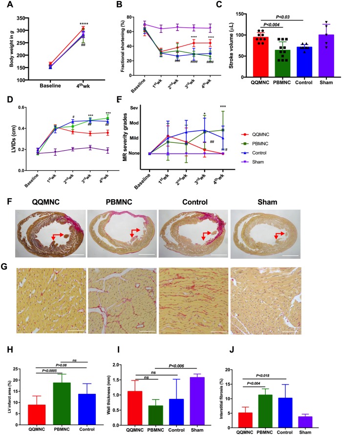
Fig 3. Echocardiographic parameters improved after four weeks in QQ-Tx group through decreasing fibrosis composition.
(A) QQ-Tx group animals gained appreciable body weight in 4 weeks. (B) Fractional shortening was notably increased from the second week after onset of MI in QQ-Tx in comparison with PB-Tx and Control littermates. (C) Stroke volume was increased and (D) left ventricular systole dimension was decreased in QQ-Tx group. (E) Mitral regurgitation events were preserved in QQ-Tx. (F and G) Representative picrosirius red staining of QQ-Tx, PB-Tx, Control, and Sham-operated rat cardiac tissues at day 28 after onset myocardial infarction. (H and I) QQ-Tx group showed preserved left ventricular infarcted area and wall thickness while PB-Tx and Control groups showed extended infarction as well as wall thinning. (J) QQ-Tx showed reduced interstitial fibrosis. LV-left ventricle. ****P<0.0001 vs. PBMNC transplanted group; #P<0.05; ###P<0.001; ####P<0.0001 vs Control group. Statistical significance was determined using 2-way ANOVA followed by Tukey’s multiple comparisons test. Results are presented as mean ± SEM.

