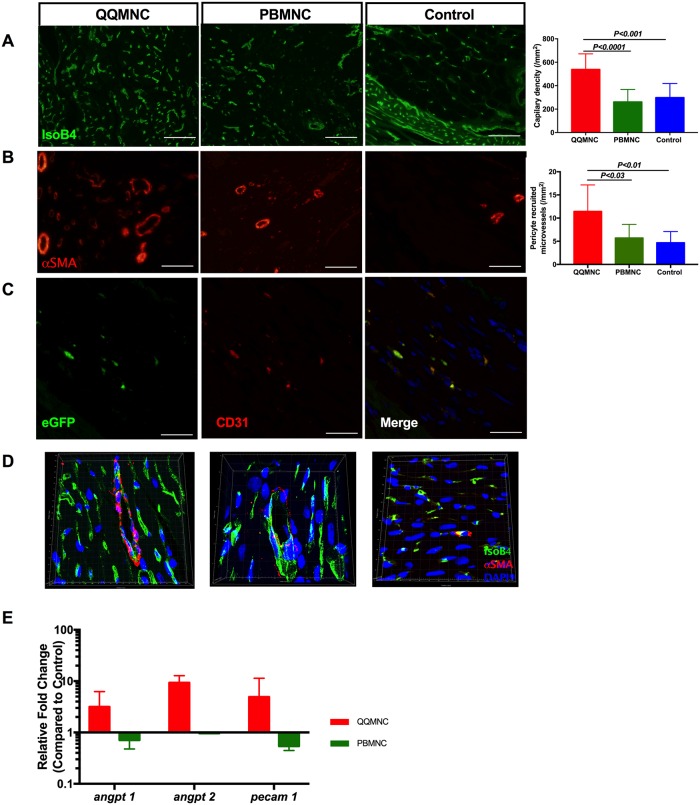
Fig 5. RACs promoted angiogenesis and arteriogenesis in myocardial infarcted area.
Six randomly detected fields were evaluated, including border and infarcted areas, with 20x magnification to obtain an average value of vascularization. (A) QQ-Tx group showed significantly increased functional capillary density and (B) αSMA stained arterioles per/mm2 whereas PB-Tx and Control groups did not. (C) QQ conditioned eGFP expressing CD31+ cells extended capillary length with incorporation into host tissues. (D) Representative Z-stack 3D construction images of Isolectin B4 and αSMA stained infarcted tissues of QQ-Tx, PB-Tx, and Control groups. (E) At day 6 post-MI, infarcted tissues of QQ-Tx showed elevated angiogenesis-related gene expression (all values are log transformed). Scale bar: 20 μm. Statistical significance was determined using Kruskal-Wallis followed by Dunn’s multiple comparisons test. Results are presented as mean ± SEM.

