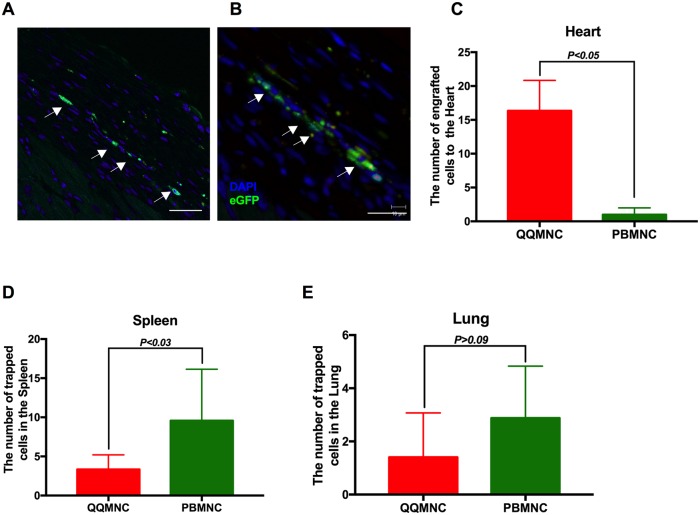
Fig 6. QQMNCs homed myocardial infarcted tissues.
(A) Representative figures of IHC cell tracking of transplanted eGFP positive QQMNCs recruited into the peri-infarcted and (B) infarcted area. (C) A significant number of eGFP positive QQMNCs homed to the infarcted area at 4 weeks in the QQ-Tx but not in the PB-Tx group. (D and E) Majority of transplanted eGFP PBMNC cells were trapped in the spleen (D) and lung tissues (E). Scale bar: 20 μm (A) and 40 μm (B). Statistical significance was determined using Mann-Whitney test. Results are presented as mean ± SEM.

