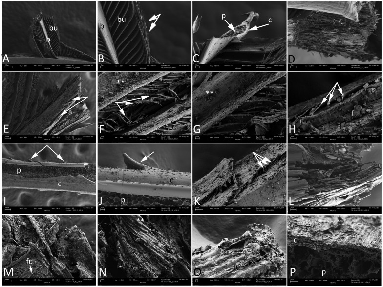
Fig 2. Scanning electron micrographs (SEM) of experimental (A-L), and Yellowstone (M-P) feathers.
A-D) barbs arising from a rachis at low (A) and higher (B-D) magnifications. Feather structure is virtually unaltered, and both barbs (b) and barbules (bu) can be seen. In Fig 2B, hooklets are seen arising from distal bars (arrows). C) Internal regions of a barb, with cortex (c) and pith (p) clearly discernible. D) Highly fibrous region of what is interpreted to be the distal rachis. E-H represent the condition 1 feathers. Loss of integrity is more obvious than in LM. E) Fraying of the rachis (arrows) reveals fibrous structure. F) Twisted and compressed barbules (arrows) and debris on the rachis and barbs (**). G) Higher magnification of rachis and barbs, with debris (**) that may be from the burial sands, or from degrading keratin flakes. H) Bent and twisted barbules (arrows) with presumed keratinous flakes (f) on the surface of feather structures. Panels I-L show the microstructural integrity of the condition 2 (350°C) feather. I) Rachis, with smooth external cortex (c) and internal pith (p). Barbs are seen arising from the surface (arrows). J) Higher magnification image showing pith (p) and cortex (c), but the cortex demonstrates thin cracks in the surface. A curved barb is still attached (arrow). K) Compressed barbules (arrows) arising from flattened barbs.; debris can be seen across the surface of these feather structures. L) Highly fibrous region of the condition 2 feather, very similar in structure to that seen in the control (D). Panels M-P show the three dimensional, coated structure of the silicified coot feather. M) Fibrous structure and overlapping barbs in low magnification, with evidence of fungal hyphae (fu) interspersed throughout. N) Region of overlapping barbs, with thin mineral coating (**). O) Thin mineral coating on the barbs in higher magnification (**); silicified fungal hyphae can also be seen (fu). P) Feather at higher magnification, revealing a fibrous outer cortex (c), and altered pith (p) interior to the cortex. Scale bars: A, E, I, M are 100 μm; K, N, P = 20 μm; O = 3 μm; all others = 10 μm.

