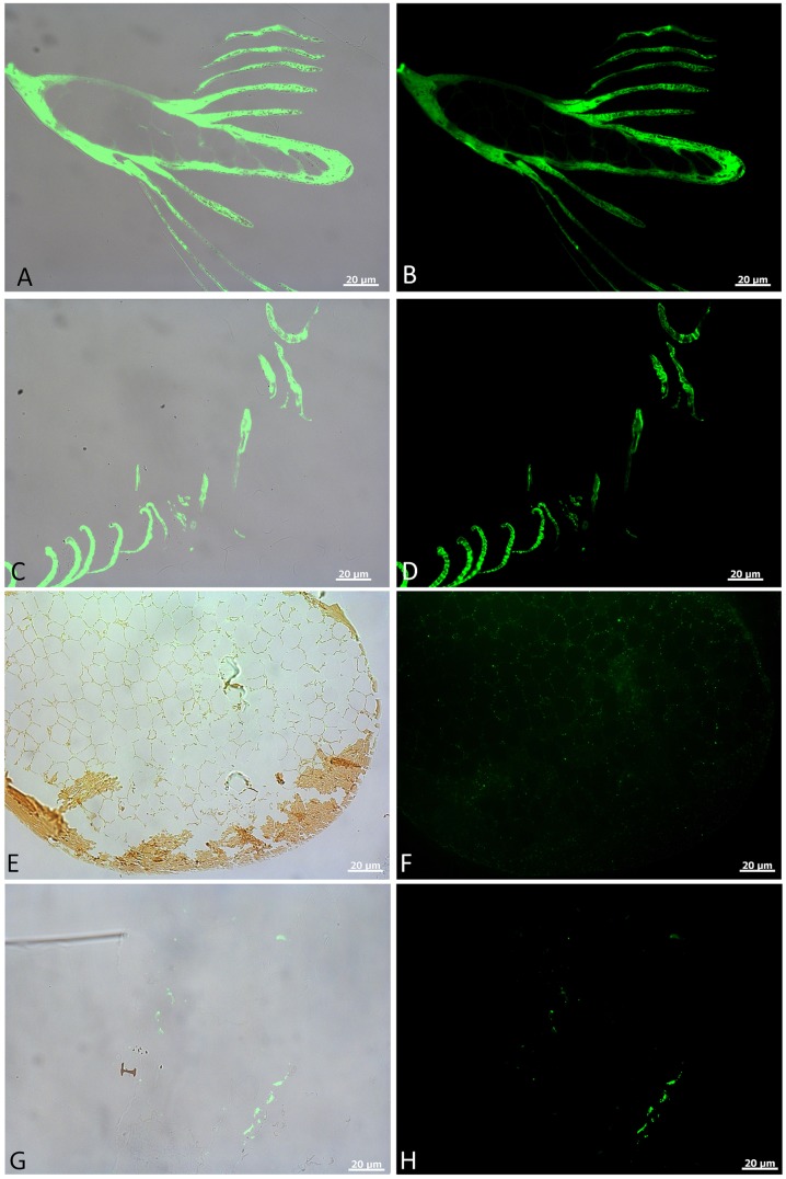
Fig 3. In situ immunofluorescence on feather tissues.
A, C, E, and G are overlay images; B, D, F, and H are fluorescence images, showing localized binding of antiserum raised against modern feathers to these experimental feathers. A, B) show in situ binding of the serum to feather rachis and barbs in control feathers. Antibody-antigen (ab-ag) complexes are demonstrated by localized green signal under fluorescent light. C, D) Virtually undiminished binding of antibodies to the condition 1 feather barbs. No spurious binding is seen on the embedding polymer, and ab-ag complexes are specific to feather structures. E, F) Cross section of a feather barb from condition 2. A thin cortex can be seen, with very thin rami of pith in E). F) Weak, but highly localized binding of antiserum to feather structures, with no binding observed outside of the tissues. G, H) Localization of ab-ag complexes to the surface of tissues seen in the Yellowstone feather. Binding is restricted to feather structure, as can be seen in G, but is intermittent and, although structurally preserved, not all feather material binds this antiserum.

