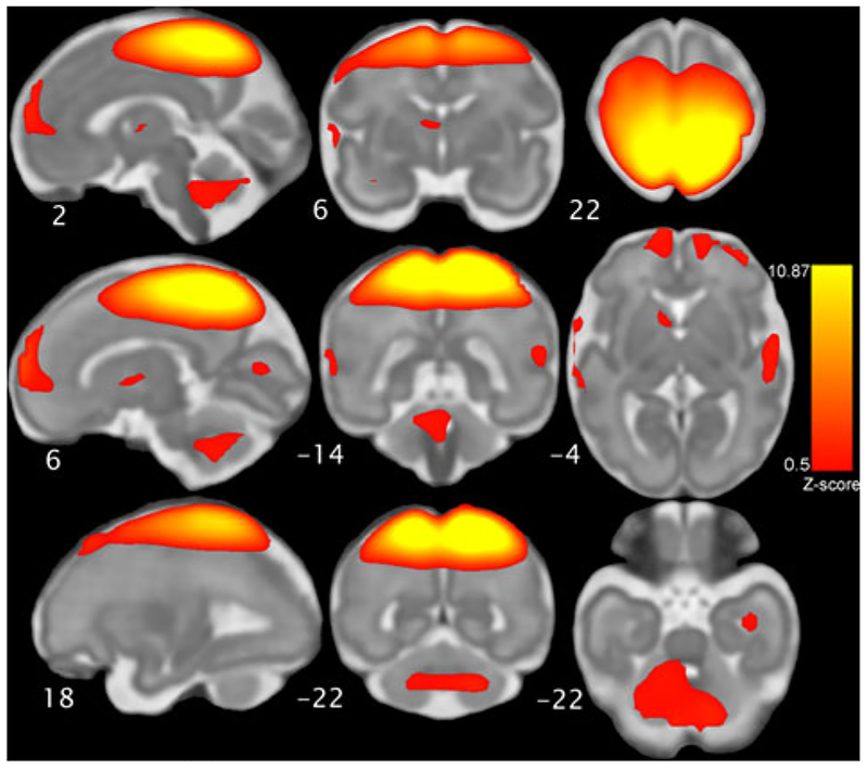
Figure 1.
The fetal sensorimotor resting state network, which was derived from group independent components analysis of resting state scans obtained in 96 fetuses. The network component map pictured here is threshold at Z = 0.5 and displayed on a 32-week gestational age template for anatomical reference. The network includes motor and sensory cortices, cerebellum, striatum, thalamus, and bilateral insula. Neurological convention is used.

