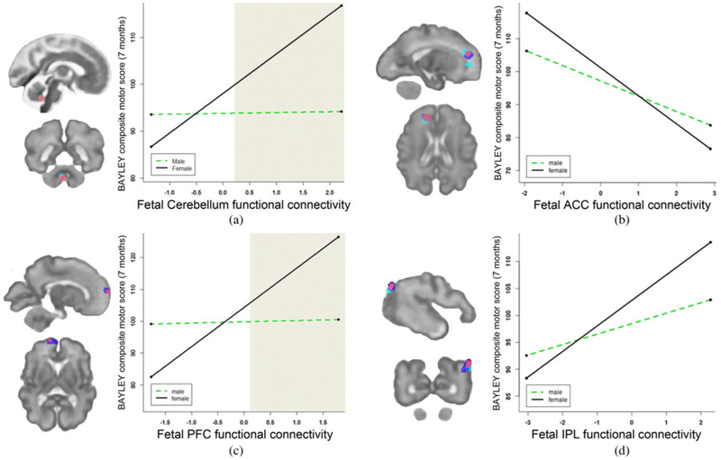
Figure 3.
Sex differences were observed in four brain regions exhibiting associations between fetal functional connectivity and Bayley motor performance at 7 months postnatal age. Mean functional connectivity values are plotted by median Bayley performance and by sex, and given for (a) the cerebellum, (b) the anterior cingulate (ACC), (c) the prefrontal cortex (PFC), and (d) the inferior parietal lobule (IPL). Structural equation models controlling for maternal education, income, anxiety, and motion during magnetic resonance imaging demonstrate significant sex interactions in the cerebellum, p = .03, and the PFC, p = .002. An examination of the region of significance in sex interactions showed these effects to be significant at higher fetal functional connectivity values (see shaded regions in [a] and [c]). When groups were tested individually, brain–behavior associations were significant among girls (ps < .01), but not boys (ps ≥ .3), across all four areas (see Table 4). Brain images to the left of plots highlight areas from which functional connectivity is plotted, corresponding to main effects of Bayley motor outcome (dark blue), interaction of Motor × Sex (cyan), and the conjunction of these (fuscia) at p < .05.

