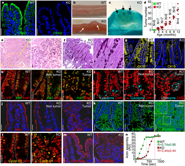
Figure 5. Intestinal adenoma development by CRAD KO.
a, CRAD expression in the small intestine. Immunohistochemistry (IHC) of mouse intestine. CRAD KO mouse serves as a negative control.
b and c, Intestinal adenoma development in CRAD KO mice. The adenomas in the small intestine of CRAD KO mice (3mo of age; b). Methylene blue staining (c). Arrows indicate intestinal adenoma.
d, Age-dependent intestinal adenoma development in CRAD KO mice. N values indicate the number of mice. Error bars: mean ± S.D. The experiment was performed once.
e, Hematoxylin and eosin (H&E) staining of intestinal adenoma (CRAD KO).
f, Periodic Acid-Schiff (PAS) staining of intestinal adenoma in CRAD KO mice.
g, Disruption of epithelial cell integrity. Cytokeratin 19 (CK19). Arrows: Villi not expressing CK19.
h, Cell hyperproliferation in CRAD KO small intestine. CRAD KOKi67.
i, Abnormal differentiation of IECs by CRAD KO. WT and CRAD KO small intestine were immunostained with Lysozyme.
j, Disorganized cell adhesion in CRAD KO mice. Cells were stained with Villin.
k, The increase of β-catenin in CRAD KO tumor.
l, Upregulation of β-catenin target genes in the intestinal adenoma of CRAD KO mice. IHC for Cyclin D1.
m, Disorganized actin cytoskeleton in CRAD KO-induced tumor. F-actin was visualized by Phalloidin staining.
n, The decrease of the actin polymerization in CRAD KO mice. Cell extracts from the small intestine were analyzed for actin polymerization assays. n=3 independent experiments. R values indicate the velocity of actin assembly.
Representative images of three independent mice per group (WT vs. KO); AU: arbitrary unit; Scale bars indicate 20μm.

