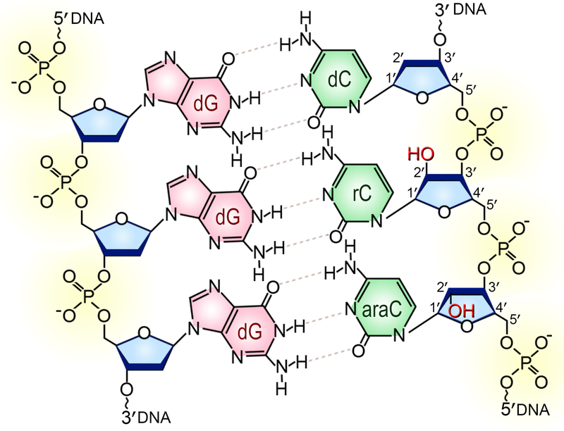
Figure 1. Chemical structure of double-stranded DNA formed by the covalently linked sugar rings (shown in blue), phosphate groups (yellow) and nitrogenous bases.
The segment shown on the diagram consists of three cytosine (green) / guanine (pink) Watson-Crick base pairs where two deoxycytidines (dC) are replaced with either cytidine (rC) or 1-β-D-arabinofuranosylcytosine (araC). The 2′-OH groups (highlighted in red) of the ribonucleotide and arabinofuranoside are on the opposite sides of the plane of the sugar. A color version of the figure is available online.

