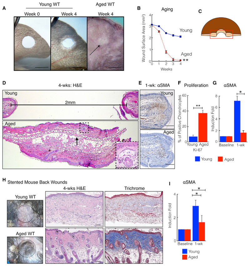
Figure 1. Aging Promotes Skin Tissue Regeneration in C57/BL6 Mice.

(A) Aged mice close ear holes to a smaller size. Shown are representative photographs of young WT and aged WT ears. The black arrow marks the healed hole.
(B) Ear hole measurements. n = 15. **p = 0.003.
(C) Schematic of histology orientation.
(D) Aged mice heal ear holes with original tissue architecture, including cartilage, subcutaneous fat, and hair follicles: H&E staining. The horizontal line measures the distance between cartilage end plates. The box taken from the dotted square illustrates a regenerated hair follicle and sebaceous gland.
(E) αSMA (brown cells) immunostaining of ears from young and aged mice. n = 10.
(F) Ear tissue from injured aged mice demonstrates increased chondrocyte proliferation and decreased fibrosis. Shown are Ki-67-expressing chondrocytes 4 weeks post-injury. n = 4. **p = 0.002.
(G) Relative αSMA transcript levels in ear wound edge tissue 1 week post-injury. n = 8. *p < 0.03.
(H) Silicone-stented back wounds on aged mice exhibit improved tissue regeneration and diminished scar formation. Shown are representative photographs, H&E staining, and trichrome staining 4 weeks post-injury. Dark blue collagen represents normal skin; pale light blue collagen represents scar. n = 5.
(I) Relative αSMA transcript levels in back wound edge tissue. n = 5. *p = 0.02.
N, biological replicates per group. Error bars are SEM. Scale bars, 100 μM (histology) and 2 mm (photographs). See also Figure S1.
