
Figure 2. Injured Young Keratinocytes Secrete SDF1 to Promote Scar Formation.
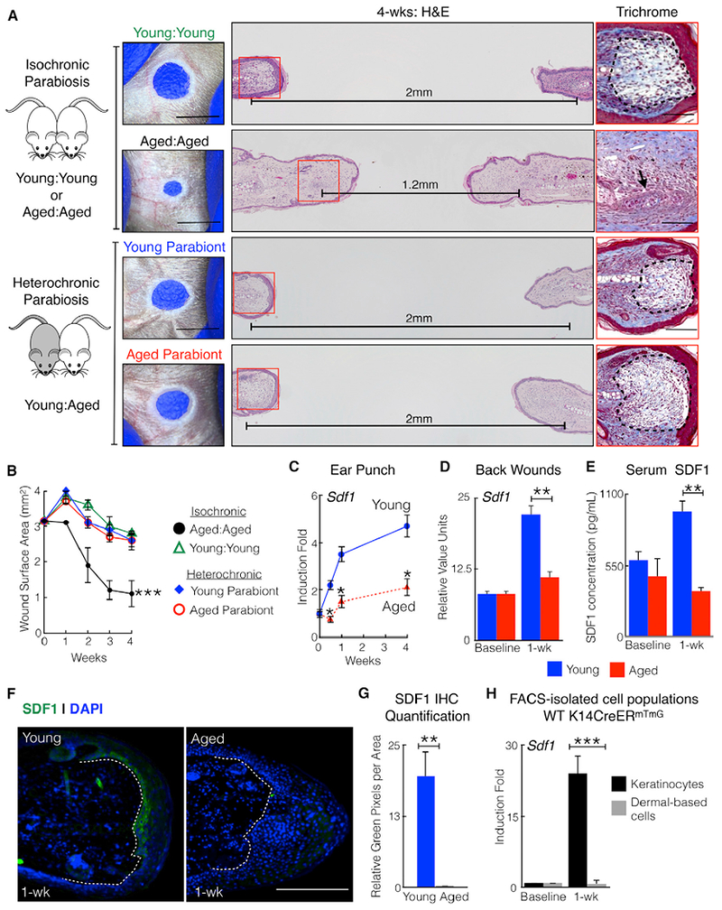
(A) A circulating factor promotes scar formation in aged mice. Shown is a schematic of parabiosis pairs, photographs, H&E staining, and trichrome staining. The trichome images are taken from the red squares. Dotted areas mark scars. The black arrow marks chondrocyte proliferation. Horizontal lines indicate the distance between cartilage end plates. n = 5.
(B) Ear hole measurements of individual parabionts. n = 5. ***p < 0.001, comparing aged: aged with young: young or either parabiont of young: aged.
(C) Mice exhibit age-dependent SDF1 induction in ear and back wounds. Shown is the relative SDF1 transcript in ear wound edge tissue of young or aged WT mice. n = 24. *p < 0.03.
(D) Relative SDF1 transcript in back skin wound edge tissue of young or aged WT mice. n = 6. **p = 0.004.
(E) Ear injury induces SDF1 blood serum levels in young but not aged mice. Shown are SDF1 blood serum levels at baseline and 1 week after ear punch injury. n = 6. **p = 0.002.
(F) Ear hole injury induces SDF1 expression in injured keratinocytes. Shown is SDF1 (green) immunostaining of ears from young and aged mice. Dotted lines identify the epidermal-dermal border. The hole is located to the right of the section. n = 6.
(G) Quantification of SDF1 immunostaining. n = 6. **p < 0.01.
(H) Relative SDF1 transcript in fluorescence-activated cell sorting (FACS)-isolated keratinocytes and dermally based cells in young mice. n = 6. ***p < 0.001.
N, biological replicates per group. Error bars are SEM. Scale bars, 100 μM (histology) and 2 mm (photographs). Nuclei are counterstained blue. See also Figure S2.