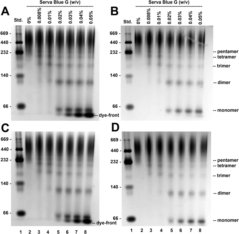
Fig. 4. Oligomeric state of MBP-5-HT3A-ICD analyzed by pCN-PAGE using a NuPAGE™ 4–12% Bis-Tris gel (Invitrogen).
6 μg (A, B) or 3 μg (C, D) of purified MBP-5-HT3A-ICD was resolved on a pCN-PAGE in the absence (lane 2) and presence of increasing concentrations of Serva Blue G as indicated in figure (lanes 3–8). Soluble proteins of the high molecular weight (HMW) kit were applied to lane 1. The gels were first stained with Biorad Bio-Safe™ Coomassie G-250, imaged (A, C), and then washed with a destaining solution (25% (v/v) methanol, 10% (v/v) acetic acid) followed by re-staining with Coomassie R-250 (B, D). Lane scan densitometry provided in Fig. 5. All gel images were captured in gray color using Gel Doc EZ Imager (Biorad).

