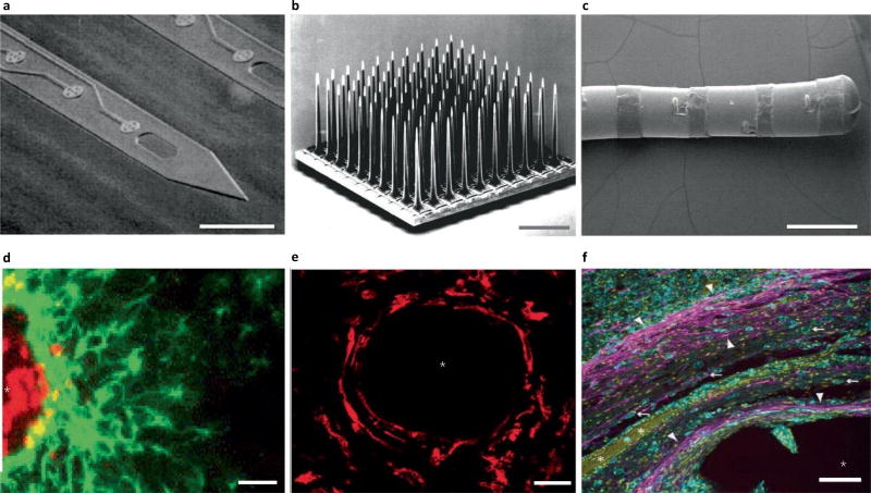
Fig. 1.
Traditional electrode arrays incite gliosis. a–f, Devices (a–c) are shown above the associated histology images (d–f). a, Michigan-style array217. b, Utah-style array218. c, DBS lead219. d, Rat histology from a Michigan-style multielectrode array (four weeks), with labelled astrocytes (GFAP, green) and microglia (ED1, red)80. e, Histology from a primate with Utah array implanted, with microglia labelled (IBA1, red)65, at 17 weeks. f, Human DBS lead implant at ~38 months, with labelled astrocytes (GFAP, magenta; white arrowheads), microglia (IBA1, cyan; white arrows) and all cell nuclei (CyQUANT, yellow)121. Scales bars: a,d,f, 100 µm; b, 1 mm; c, 2 mm; e, 28 µm. The asterisks in d–f indicate injury. ED1, antibody to cluster of differentiation 68; IBA1, ionized calcium binding adaptor molecule 1. Figure reproduced from: a, ref.217, IEEE; b, ref.218, Elsevier; c, ref.219, Oxford Univ. Press; d, ref.80, Elsevier; e, ref.65, IOP Publishing; f, ref.121, Springer.

