Fig.5.
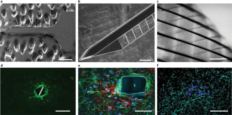
Next-generation arrays mitigate gliosis. a–f, Devices (a–c) are shown above the associated histology images (d–f). a, A mechanically adaptive nanocomposite microelectrode becomes compliant on implantation163. b, A hollow-architecture parylene-based microelectrode places sites away from the stiff penetrating shaft, along 4-µm-wide lateral support arms148. c, A syringe-injectable mesh electronics mimics brain parenchyma with sites featured along an interwoven structure172. d, Astrocytes labelled (GFAP, green) around mechanically compliant probe at eight weeks161. e, Astrocytes (GFAP, red), microglia (OX42, green), and all cells (Hoechst, blue) labelled around the stiff electrode-penetrating shaft (S) and lateral edge (L) at four weeks148. f, Astrocytes labelled (GFAP, cyan) around a syringe-injected mesh (blue) at one year171. Scale bars: a, 500 µm; b,d,f, 100 µm; c, 250 µm; e, 50 µm. Figure reproduced from: a, ref.163, IOP Publishing; b,e, ref.148, Elsevier; c, ref.172, American Chemical Society; d, ref.161, IOP Publishing; f, ref.171, Nature America Inc.

