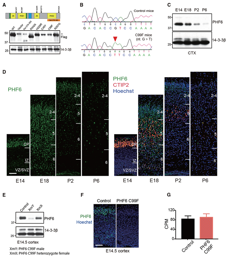
Figure 1. Generation of Patient-Specific PHF6 Mutant Mouse Model of BFLS.

(A) Top: schematic of PHF6 protein structure. Dots: patient-specific mutations in BFLS; nucleus localization sequence (green);nucleolar localization sequence (blue). Bottom: immunoblot of lysates of 293T cells transfected with expression plasmids encoding FLAG-tagged PHF6, patient-specific mutations of PHF6, or control vector using an antibody to FLAG or 14-3-3β, the latter serving as loading control. Non-specific immunoreactive band (*).
(B) Sequencing traces of wild-type and C99F PHF6 alleles.
(C) Immunoblot of PHF6 in lysates of the cerebral cortex in mice at embryonic day 14 (E14), E18, postnatal day 2 (P2), and P6.
(D) Immunohistochemical analyses of PHF6 (left) and CTIP2 (right) in sections of the cerebral cortex from E14, E18, P2, and P6 mice. DNA dye bisbenzimide (Hoechst) was also used (right). CP, cortical plate; IZ, intermediate zone; VZ/SVZ, ventricular zone/subventricular zone. Scale bar, 50 μm.
(E) Immunoblot of PHF6 in lysates of the cerebral cortex from control male, C99F male, and C99F heterozygote female mice.
(F) Immunohistochemical analyses of PHF6 in sections of the cerebral cortex from E14.5 control and C99F mice. Hoechst was also used. Scale bar, 50 μm.
(G) The levels of PHF6 mRNA in the cerebral cortex of control and C99F mice in RNA-seq analysis (n = 5). CPM, counts per million.
Data are presented as means ± SEMs. See also Figure S1 and Table S1.
