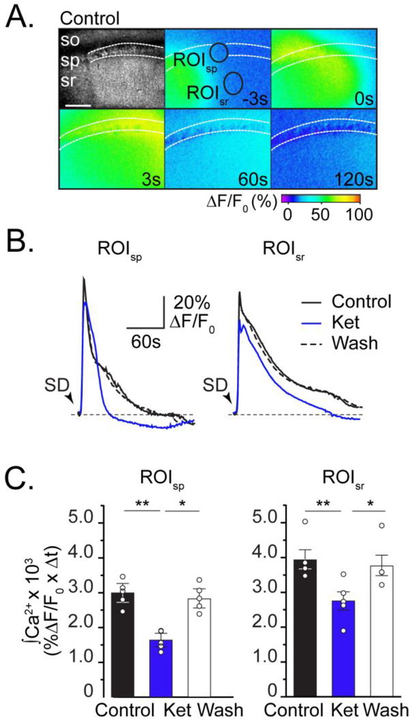
Figure 3. Ketamine reduces neuronal intracellular Ca2+ accumulation during SD.
A: Top left panel: Transmitted light image, showing stratum oriens (so), stratum pyramidale (sp), stratum radiatum (sr) in area CA1. Pseudo colored images show GCaMP5G fluorescence collected during SD in control conditions. Numbers in each frame indicate time (in seconds) in relation to peak Ca2+ during SD, and black circles are regions of interest surrounding predominately pyramidal cell bodies or dendrites in stratum pyramidale (ROIsp )or radiatum (ROIsr), respectively. Scale bar = 100μm. B: Data extracted from ROIsp and ROIsr show that Ca2+ transients during SD in ketamine (blue) recover faster than control (black), and are reversible after ketamine wash out (dashed). Black arrowheads indicate SD onset. C: Summary data (n=5), show that ketamine reversibly reduces total neuronal Ca2+ accumulation in both ROIs (integrals of 120s transients; see Methods). *P<0.05, **P<0.01

