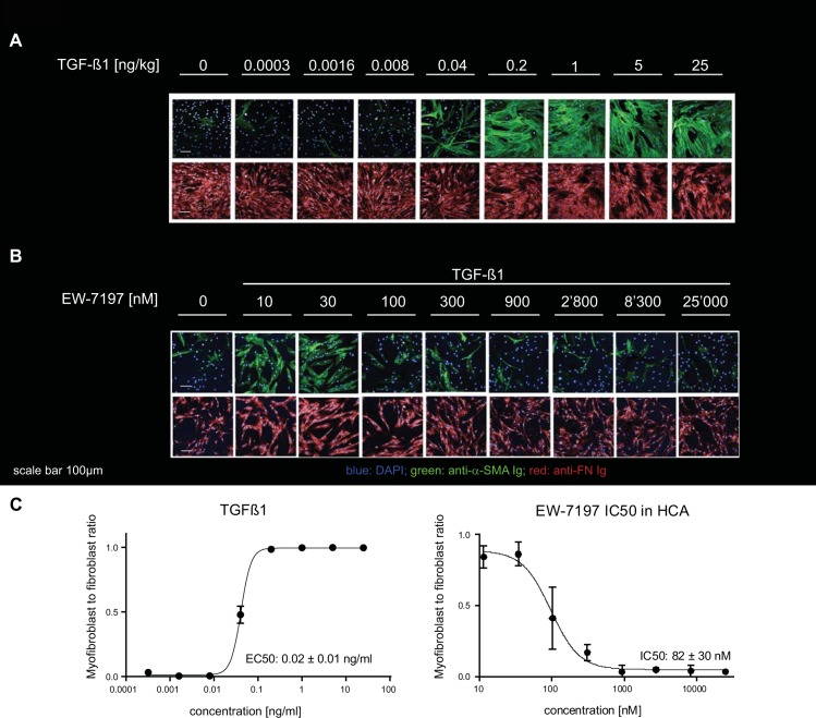
Fig 1. TGF–β1–stimulated normal human lung fibroblast–to–myofibroblast differentiation and inhibition thereof by using the ALK5 inhibitor EW–7197.
(A) Effect of increasing concentrations of TGF–β1 ranging from 0–25 ng / ml on NHLF fibroblasts as captured by high–content confocal microscopy. (B) The effect of TGF–β1 (5 ng / ml) is inhibited by increasing concentrations of the ALK5 inhibitor EW–7197 from 10 to 25,000 nM concentration. 0 panel had no TGF–β1. (C) Concentration response curves of TGF–β1 and EW–7197 in presence of 5 ng / ml TGF–β1 were generated from the myofibroblast to fibroblast ratios as classified by the trained SVM. Curves were generated from data shown in panels A, B (n = 2). EC50 and IC50 values represent mean ± SD (n = 7). Scale bar 100 μm.

