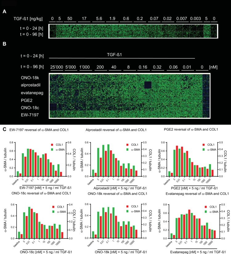
Fig 6. Reversal of TGF–β1–induced NHLF myofibroblast phenotype.
(A) After starvation NHLF cells were stimulated with a dilution series of TGF–β1 (0.003–50 ng / ml) either for 24 h (after which cells were washed and cultured for further 72 h without TGF–β1, top row) or for the entire duration of 96 h (bottom row). Cells were fixed and immunostained to detect the myofibroblast markers FN and α–SMA by high–content confocal microscopy. Shown are the green channel images representing α–SMA. The final assay concentration of DMSO was 0.6% in all wells. (B) Confocal images of NHLF cells that were differentiated for 24 h with 5 ng / ml TGF–β1 into myofibroblasts. The cells were then washed 3 times to remove TGF–β1 and incubated for 72 h with increasing concentrations (0–10,000 nM) of the agonists ONO–18k, alprostadil, evatanepag, PGE2 and ONO–18c, as well as with the ALK5 blocker EW–7197. (C) Data represent α–SMA (green bars) and COL1 (red bars) quantified by MS / MS and normalized to tubulin, at t = 96 h after TGF–β1 addition. Number of sample (n = 1) is shown in each figure.

