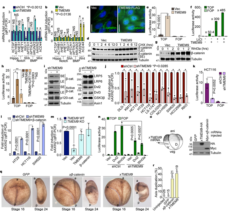
Fig. 2. Activation of Wnt/β-catenin signaling by TMEM9.
a and b, Screening of cell signalings. mRNA expression in CRC cells (a) and IECs (b) was analyzed by qRT-PCR. c-d, Upregulation of β-catenin by TMEM9. IF staining of β-catenin (c). HeLa cells were analyzed for β-catenin protein half-life using cycloheximide (CHX; 100μg/ml; d). e and f, Activation of the β-catenin reporter by TMEM9. CCD-841CoN cells were analyzed by β-catenin reporter assays (e). 293T cells were transfected with each plasmid and treated with LiCl (25mM, 24hr; f). g, Enhancement of β-catenin stabilization by TMEM9 upon Wnt3a. IB of 293T cells transiently expressing Ctrl or TMEM9 upon Wnt3a treatment (200ng/ml). h, Decreased TMEM9-activated β-catenin reporter by iCRT14. 48hr after overexpression of TMEM9, CCD-841CoN cells were incubated with vehicle or iCRT14 (100μM, 12hr). i, Inhibition of β-catenin by TMEM9 depletion. IB of HCT116 (shCtrl vs. shTMEM9). j and k, Decreased β-catenin transcription activity by shTMEM9. qRT-PCR of AXIN2 (j) and luciferase activity (k). l and m, Rescue of Wnt/β-catenin activity by ectopic expression of β-catenin in TMEM9-depleted CRC. CRC cells were transfected with indicated plasmids for 24hr (l). HCT116 (TMEM9 WT vs. KO) cells were stably transduced with either TMEM9 or β-catenin (m). n, Wnt/β-catenin signaling activation by TMEM9 at the downstream of Dvl2 and Wnt3a. HCT116 cells were co-transfected with β-catenin reporter plasmids and Dvl2 or treated with Wnt3a for luciferase assays. o-r, In vivo activation of Wnt/β-catenin signaling by xTMEM9 in frog embryos. Xenopus laevis embryos were injected with each mRNA into ventral-vegetal blastomeres at the 4-cell stage (o). Expression of microinjected Myc-xβ-catenin or xTMEM9-HA mRNA was confirmed by IB (p). Axis duplication was analyzed at the neural fold (st16) and the tail buds (st24) stages (q). Quantification of axis duplication (r). ani: animal pole; veg: vegetal pole; V: ventral region; D: dorsal region; a: anterior; p: posterior; a’: secondary anterior axis. n: biologically independent samples. Experiment was performed once. Images in c and blots in d, g, I and p are representative of three independent experiments with similar results; Scale bars=20μm; NS: Not significant; Error bars: mean ± S.D.; Two-sided unpaired t-test.

