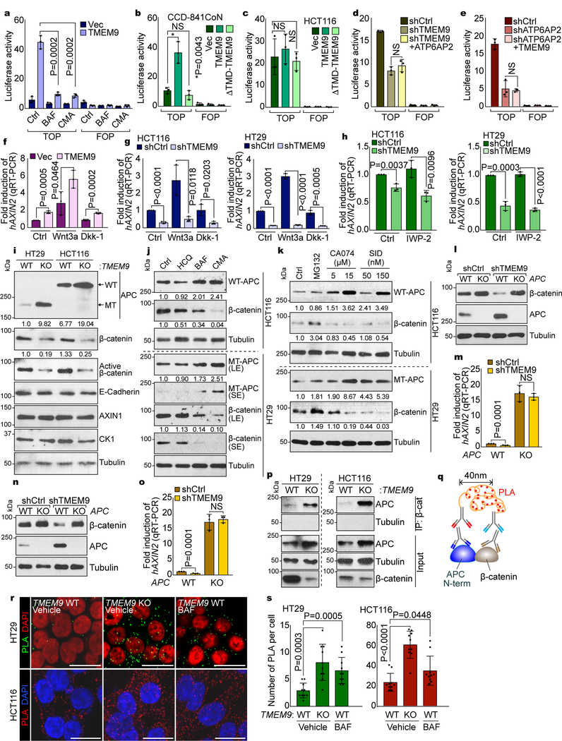
Fig. 4. Decrease of APC by TMEM9-induced v-ATPase activation.
a, Suppression of TMEM9-activated β-catenin reporter by v-ATPase inhibitors. 239T cells were transfected with β-catenin reporter plasmids and treated with BAF or CMA for 24hr. b and c, Requirement of TMEM9-TMD for TMEM9-induced β-catenin reporter activation. CCD-841CoN (b) and HCT116 (c) cells were transfected with indicated plasmids and analyzed by luciferase assays. d and e, No effect of ATP6AP2 and TMEM9 on β-catenin reporter activity in TMEM9- or ATP6AP2-depleted CRC cells, respectively. shCtrl, shTMEM9 (d), or shATP6AP2 (e) plasmids were co-transfected with ATP6AP2 (d) or TMEM9 (e) plasmids, respectively. f and g, Increased β-catenin transcription activity by TMEM9 independently of Wnt agonist or antagonist. IECs (f) and CRC cells (g) were incubated with Wnt3a (50ng/ml) or Dkk-1 (100ng/ml) for 12hr and analyzed by AXIN2 qRT-PCR.h, Downregulation of Wnt/β-catenin signaling by shTMEM9 independently of Wnt ligand secretion. After transfection, cells were incubated with IWP-2 (2μM) for 12hr. qRT-PCR of AXIN2. i, Upregulated APC protein by TMEM9 depletion. TMEM9 WT and KO cells were analyzed by IB. j, Increased APC protein by v-ATPase inhibition. HCT116 and HT29 cells were incubated with indicated reagents (HCQ: 25μM; BAF: 3nM; CMA: 0.3nM) for 6hr. IB analysis and quantification using ImageJ.k, APC upregulation by the inhibition of lysosomal protein degradation. CRC cells were incubated with Cathepsin inhibitors (CA074 and SID26681509) for 12hr. IB and quantification using ImageJ. l-o, Loss of TMEM9 depletion-downregulated Wnt/β-catenin signaling by APC KO. HCT116 (l and m) and HT29 (n and o) cells were analyzed for IB and qRT-PCR of AXIN2. p-s, The increased interaction between APC and β-catenin by TMEM9 KO. Co-IP analysis (p). Scheme of Duolink assay monitoring APC-β-catenin binding (q). Cells were incubated with APC-N terminal (mouse) and β-catenin (rabbit) antibody followed by reaction with PLA probes. Dots indicate an interaction between APC and β-catenin (r). Protein interaction was quantified (s; n=10 independent samples) using manufacturer’s software (Sigma). Representative images of three experiments with similar results; Scale bars=20μm; NS: Not significant; Error bars: mean ± S.D. from n=3 independent experiments, except from s; Two-sided unpaired t-test.

