
Figure 2. MIF overexpression in Mif-lung Tg2.1 founder mice and regulation transcription by The MIF −794 CATT5–8 promoter polymorphism in response to β-glucan in a CATT-length dependent manner.
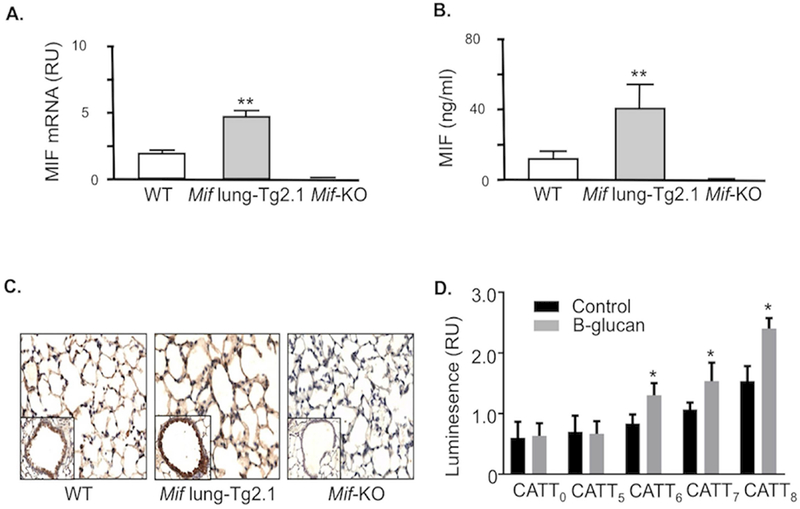
(A) Quantitative PCR analysis of Mif mRNA in lung tissue harvested from 3 representative wild type (WT), Mif lung-Tg2.1, and Mif-KO mice at 6 weeks of age. (B) ELISA analysis of MIF content in bronchoalveolar lavage fluid obtained from three mice of indicated strains. (C) Immunohistochemistry analysis of mouse lung tissue stained with anti-MIF pAb showing prominent staining in alveolar epithelium and increased MIF content in Mif lung-Tg2.1 mice. Tissue sections from a Mif-KO mouse lung was used as an antibody specificity control (200×). Insets show a representative terminal bronchus and sections are representative of three mice per genotype. **p<0.01 by Student’s t test, two tailed. (D) Human pulmonary A549 cells transfected with MIF promoter/luciferase reporter fusion plasmids bearing 0, 5, 6, 7, and 8 CATT repeats were stimulated with 100 ng/ml β-glucan for 8 hrs and luciferase expression expression measured as luminescence relative units (RU). Mean ± SD for quadruplicate determinations, replicated twice. *p<0.05 for comparisons to 5-CATT or to β-glucan stimulation versus control by Student’s T test (two-tailed).